Simulation of Cellular Traction Force Based Deflection of PDMS Micropillars
Cells are complex entities which not only passively sense external stimuli (viz. chemical, optical or mechanical) but also interact with extracellular matrix (ECM) by regulating cellular behavior such as growth, proliferation, migration, etc. Monitoring cell growth and migration of adherent cells becomes a crucial factor in determining cell-cell and cell-substrate interaction, important for formation of tissues, signal transduction pathways and body’s defense system. The detection of such interactions can serve as unique index to probe trivial changes in single cells prior to the time when physiological and morphological changes can be resolved. This kind of research would have profound influence and application in physiology, medicine and cell biology fields.
Highly complaint arrays are used to study the deflection of the tips of microfabricated micropillars which is directly proportional to cell-generated traction forces during cell migration and contraction by various groups. Present study discusses the effect of such cellular traction force over different geometries and varying elasticity of PDMS micropillars.
Numerical modeling of the deflection of PDMS micropillars have been accomplished using COMSOL Multiphysics® software. The Solid Mechanics interface was used to model circular micropillar structures for varying diameter from 5-50 µm with constant gap of 10 µm. Variation in Young’s modulus of PDMS corresponding to different base and curing agent ratio of 5:1 - 20:1 were also employed for the above geometries. Distributed force in pN range corresponding to epithelial cells was applied near top edges of the micropillars to compute total deflection, stresses and strain on the micropillars.
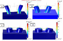
Simulation results showing deflection of micropillar tip were observed in different diameters of circular micropillar. Figure 1 shows the images of PDMS micropillar deflection and stress for pillar diameter 5 µm and 10 µm simulated with higher young’s modulus of 3.72MPa corresponding to 5:1 PDMS composition. The simulated stress was observed to be maximum for micropillar of 5 µm diameter for all PDMS compositions and maximum deflection of micropillar was observed for 5 µm and 10 µm diameter for all PDMS compositions as shown in Figure 2.
COMSOL Multiphysics® was successfully used to study the deflection of micropillar tips induced due to cell-generated traction force. Simulation results show appreciable deflection for smaller micropillar geometries with 5 and 10 µm diameters as compared to higher geometries. Furthermore, PDMS composition of 20:1 showed maximum deflection due to lower young’s modulus. The above simulated results would thus help in further designing of actual PDMS based micropillar arrays for actual measurement of micropillar deflection to study cellular biomechanics.
ダウンロード
- wala_poster.pdf - 1.33MB
- wala_abstract.pdf - 0.25MB